

Ultrasounds and x-rays in pancreatitis
4 imaging tests that can evaluate the structure of the pancrease
-Imaging tests can evaluate the structure of the pancreas. Scarring of the pancreas is often seen in chronic pancreatitis. Inflammation and / or swelling can be identified during active pancreatitis.
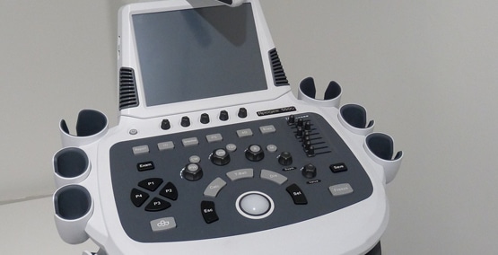
Ultrasounds
Ultrasound of the abdomen can be done in the same way an ultrasound is done to evaluate babies during a mother's pregnancy. Or it can be done as part of an endoscopy.
During the typical x-ray like procedure, sound waves are sent toward the pancreas through a handheld device that moves over the abdomen. The sound waves transmit pictures of the pancreas and the ducts that drain the pancreas. However, there are limitations to this test. Anyone overweight or who has lots of gas can affect the ability to get a clear picture of the pancreas.
During an upper endoscopy, an ultrasound can also be done. At this time it is viewed through the stomach or the upper small intestine.
CT scans
Computerized tomography (CT) is an x-ray that produces 3-dimensional images of parts of the body. It can show the extent of damage to the pancreas and if there are any gallstones or tumors.
It can be used to get more precise images of the pancreas if an ultrasound doesn't give enough detail.
MRCP
Magnetic resonance cholangiopancreatopgraphy (MRCP) is a kind of magnetic resonance imaging (MRI) to get detailed images of the pancreas without using x-rays or radiation. Its images are similar to a CT scan, using magnets instead of radiation, but it is more costly. During the procedure, dye is injected into the veins to outline the pancreas, the gallbladder and their ducts.
Bone Density
Bone strength may be evaluated by using a bone density test in chronic pancreatitis. Why? Because chronic pacreatitis can cause EPI (exocrine pancreatic insufficiency) with malabsorption of nutrients. Low levels of calcium and vitamin D can cause bone weakening so a scan of the spine and the legs for osteoporosis may be indicated.